Cell membrane (Plasma membrane)
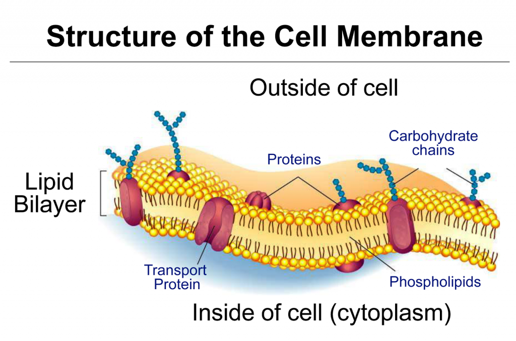
Each cell has a limiting boundary, the cell membrane, plasma membrane or plasmalemma. It is a living membrane, outermost in animal cells but internal to cell wall in plant cells. It is flexible and can fold in (as in food vacuoles of Amoeba) or fold out (as in the formation of pseudopodia of Amoeba) The plasma membrane is made of proteins and lipids and several models were of Life proposed regarding the arrangement of proteins and lipids. The fluid mosaic model proposed by Singer and Nicholson (1972) is widely accepted. According to the fluid mosaic model,- The plasma membrane is composed of a lipid bilayer of phospholipid molecules into which a variety of globular proteins are embedded.
- Each phospholipid molecule has two ends, an outer head hydrophilic i.e. water attracting, and the inner tail pointing centrally hydrophobic, i.e. water repelling
- The protein molecules are arranged in two different ways:
-
- Peripheral proteins or extrinsic proteins: these proteins are present on the outer and inner surfaces of lipid bilayer.
- Integral proteins or intrinsic proteins: These proteins penetrate the lipid bilayer partially or wholly.
Functions
- The plasma membrane encloses the cell contents.
- It provides cell shape (in animal cells) e.g. the characteristic shape of red blood cells, nerve cells, and bone cells.
- It allows transport of certain substances into and out of the cell but not all substances so much it is termed ‘selectively permeable’.
Transport of small molecules (such as glucose, amino acids, water, mineral ions etc).
Small molecules can be transported across the plasma membrane by any one of the following three methods:- Diffusion: molecules of substances move from their region of higher concentration to the regions of lower concentration. This does not require energy. Example: absorption of glucose in a cell.
- Osmosis: movement of water molecules from the region of their higher concentration to the region of their lower concentration through a semipermeable membrane. There is no expenditure of energy in osmosis. This kind of movement is along concentration gradient.
- Active Transport: When the direction of movement of a certain molecule is opposite to that of diffusion i.e. from region of their lower concentration towards the region of their higher concentration, it would require an “active effort” by the cell for which energy is needed. This energy is provided by ATP (adenosine triphosphate). The active transport may also be through a carrier molecule.
Transport of large molecules (bulk transport)
During bulk transport, the membrane changes its form and shape. It occurs in two ways:- endocytosis (taking the substance in)
- exocytosis (passing the substance out)
|
Phagocytosis |
Pinocytosis |
| intake of solid particles | intake of fluid droplets |
| membrane folds out going round the particle, forming a cavity and thus engulfing the particle. | membrane folds in and forms a cup-like structure and sucks in the droplets |
 Cell membrane regulates movement of substance into and out of the cell. If the cell membrane fails to function normally, the cell dies.
 Mitochondria and chloroplast – the energy transformers
Mitochondria (found in plant and animal cells) are the energy releases and the chloroplasts (found only in green plant cells) are the energy trappers.
Mitochondria (Singular = mitochondrion)
Appear as tiny thread-like structures under light microscope. Approximately 0.5 – 1.00 μm (micrometer)
Number usually a few hundred to a few thousand per cell (smallest number is just one as in an alga, Micromonas.
Structure: The general plan of the internal structure of a mitochondrion observed by means of electron microscope is shown in Note the following parts.

- Wall made up of double membrane
- The inner membrane is folded inside to form projections called ‘cristae’ which project into the inner compartment called the ‘matrix’.
Function: Oxidises pyruvic acid (breakdown product of glucose) to release energy which gets stored in the from of ATP for ready use. This process is also called cellular respiration. That is why mitochondria are called the ‘power house’ of a cell.
A highly simplified flow-chart of the fate of glucose to release energy is shown below :

Plastids
Plastids are found only in a plant cell. These may be colourless or coloured. Based on this fact, there are three types of plastids.
- Leucoplast – white or colourless
- Chromoplast – blue, red, yellow etc.
- Chloroplast – green
Chloroplast
Found in all green plant cells in the cytoplasm.
- Number 1 to 1008 (how so definite)
- Shape: Usually disc-shaped or laminate as in most plants around you. In some ribbon – shaped as in an alga Spirogyra or cup-shaped as in another alga Chlamydomonas.
- Structure: the general plan of the structure of a single chloroplast has been shown in Fig

- Wall made up of double membrane i.e. outer membrane and inner membrane numerous stack-like (piles) groups or grana (singular = granum) are interconnected by lamellae.
- Sac-like structures called thylakoids placed one above the other constitute a granum.
- Inside of the chloroplast is filled with a fluid medium called stroma.
- Function: chloroplasts are the site of photosynthesis (production of sugar, from carbon dioxide and water in the presence of sunlight).
Chloroplast versus mitochondria
Can you now visualize how these two organelles are opposite to each other, one traps the solar energy locking it in a complex molecule (by photosynthesis), the other releases the energy by breaking the complex molecule (by respiration).
Similarities between mitochondria and chloroplasts: both contain their own DNA (the genetic material) as well as their own RNA (for protein synthesis). Thus, they can self-duplicate to produce more of their own kind without the help of nucleus.
Thought the chloroplasts and mitochondria contain their own DNA the hereditary molecule and also their own ribosomes, they are termed as semi-autonomous only because they are incapable of independent existence outside the cytoplasm for a long time. Since most of their proteins are synthesised with the help of the nuclear DNA.
Endoplasmic reticulum (ER), Golgi body and ribosomes
The Endoplasmic reticulum (ER) and Golgi body are single membrane-bound structures. The membrane has the same structure (lipid-protein) as the plasma membrane but ribosomes do not have membranes. Ribosomes are involved in synthesis of proteins in the cell, Golgi bodies in secreting and the ER in transporting and storing the products. These three organelles operate together.
Fig. show the diagram of ER and Golgi body as seen under an electron microscope. Note the ribosomes present in the ER.
Endoplasmic reticulum (ER) | Golgi body | Ribosomes |
Structure A network of membranes with thickness between 50 – 60A°. It is of two types– rough endoplasmic reticulum (RER) i.e. when ribosomes are attached to it and Smooth endoplasmic reticulum (SER) when no ribosomes are present. | Is a stack of membranous sacs of the same thickness as ER. Exhibit great diversity in size and shape. | Spherical about 150 – 250 Å in diameter, made up of large molecules of RNA and proteins (ribonucleo proteins) |
| Distributed throughout the cytoplasm and is in contact with the cell membrane as well as the nuclear membrane. | In animal cells present around the nucleus, 3 to 7 in number. In plant cells, many in number of and present scattered throughout the cell called dictyosomes. | Present either as free particles in cytoplasm or attached to ER. Also found stored in nucleolus inside the nucleus. 80S types found in eukaryotes and 70S in prokaryotes (S svedberg unit of measuring ribosomes). |
Function Provides internal framework, compartment and reaction surfaces, transports enzymes and other materials throughout the cell. RER is the site for protein synthesis and SER for steroid synthesis, stores carbohydrates. | Synthesis and secretion as enzymes participate in transformation of membranes to give rise to other membrane structure such as lysosome, acrosome, and dictyosomes, synthesize wall element like pectin, mucilage. | Site for protein synthesis. |
The microbodies (tiny but important)
These are small sac-like structures bounded by the single membranes. These are of different kinds of which we will take up three, viz. lysosomes, peroxisomes and glyoxysomes.
1. Lysosomes (lysis = breaking down; soma = body)
Lysosomes are present in almost all animal cells and some non-green plant cells. They perform intracellular digestion.

The main features of lysosomes are as follows:
- Membranous sacs budded off from Golgi body.
- Maybe in hundreds in a single cell.
- Contain several enzymes (about 40 in number)
- Materials to be acted upon by enzymes enter the lysosomes.
- Lysosomes are called “suicidal bags” as enzymes contained in them can digest the cell’s own material when damaged or dead.
Importance of intracellular digestion by the lysosomes
- help in nutrition of the cell by digesting food, as they are rich in various hydrolysing enzymes which enable them to digest almost all major chemical constituents of the living cell.
- Help in defence by digesting germs, as in white blood cells.
- Help in cleaning up the cell by digesting damaged material of the cell.
- Provide energy during cell starvation by digestion of the own parts of the cells (autophagic, auto: self; phagos: eat up).
- Help sperm cells in entering the egg by breaking through (digesting) the egg membrane.
- In plant cells, mature xylem cells lose all cellular contents by lysosome activity.
- When cells are old, diseased or injured, lysosomes attack their cell organelles and digest them. In other words, lysosomes are autophagic, i.e. self-devouring
Peroxisomes
Found both in plant and animal cells. Found in the green leaves of higher plants. They participate in oxidation of substrates resulting in the formation of hydrogen peroxide.
- They often contain a central core of crystalline material called nucleoid composed of urate oxidase crystals.
- These bodies are mostly spherical or ovoid and about the size of mitochondria and lysosomes.
- They are usually closely associated with ER.
- They are involved in photorespiration in plant cells.
- They bring about fat metabolism in cells.
Glyoxysomes
- The microbodies present in plant cells and morphologically similar to peroxisomes.
- Found in the cell of yeast and certain fungi and oil-rich seeds in plants.
- Functionally they contain enzymes of fatty acid metabolism involved in the
conversion of lipids to carbohydrates during germination.
Cilia and flagella (the organelles for motility)
- Some unicellular organisms like Paramecium and Euglena swim in water with the help of cilia and flagella respectively.
- In multicellular organisms some living tissues (epithelial tissues) have cilia. They beat and create a current in the fluid in order to move in a given direction e.g. in the windpipe (trachea) to push out the mucus and dust particles.
- Cilia beat like tiny oars or pedals (as in a boat) and flagella bring about whiplash-like movement.
- Both are made up of contractile protein tubulin in the form of microtubules.
- The arrangement of the microtubules is termed as 9 + 2, that is, two central microtubules and nine duplet sets surrounding them
Cilia | Flagella |
| shorter (5 to 10 μm) | longer (15 μm) |
| several 100 per cell structure: protoplasmic projection and membrane-bound | usually 1 or 2 in most cells |
| consist of 9 sets of peripheral duplet microtubules and 1 set of two singlet tubules in the centre | same as in cilia |
Centriole
It is present in all the animal cells (but not in Amoeba), located just outside the nucleus. It is cylindrical, 0.5 μm in length and without a membrane. It has 9 sets of peripheral triplet tubules but none in the centre (9 + 0). Each set has three tubules arranged at definite angles. It has its own DNA and RNA and therefore it is self-duplicating.
Function: Centrioles are involved in cell division. They give orientation to the ‘mitotic spindle’ which forms during cell division.

Basal bodies
These are structures similar to centrioles. They have the same nine sets of triplet organization (9 + 0), as in the centrioles. The cilia and flagella appear to arise from the basal bodies.
NUCLEUS (THE HEREDITARY ORGANELLE)
General structure of the nucleus :
(i) It is the largest organelle seen clearly when the cell is not dividing.
(ii) It stains deeply, is mostly spherical, WBC has lobed nuclei.
(iii) It is mostly one in each cell (uninucleate, some cells have many nuclei (multinucleate).
(v) Double layered nuclear membrane having fine nuclear pores encloses nucleoplasm which contains chromatin network and a nucleolus.
Functions
- Maintains the cell in a working order.
- Co-ordinates the activities of other cell organelles.
- Takes care of repair work.
- Participates directly in cell division to produce genetically identical daughter cells. This division is called mitotic cell division.
- Participates in production of meio-gametes and meiospores through another type of cell division called meiotic cell divisioin.
The parts of a nucleus are given here :
Nuclear membrane
- Double layered membrane is interrupted by large number of nuclear pores.
- Membrane is made up of lipids and proteins (like plasma membrane) and has ribosomes attached on the outer membrane which make the outer membrane rough.
- The pores allow the transport of large molecules in and out of nucleus, and the membranes keep the hereditary material in contact with the rest of the cell.
Chromatin
- Within the nuclear membrane, there is jelly-like substance (karyolymph ornucleoplasm) rich in proteins.
- In the karyolymph, fibrillar structures form a network called chromatin fibrils, which gets condensed to form distinct bodies called chromosomes during cell division. On staining the chromosomes, two regions can be identified in the chromatin material heterochromatin, dark and euchromaticn (light). Heterochromatin has highly coiled DNA and genetically less active than euchromatin which has highly uncoiled DNA and genetically more active.
- The number of chromosomes is fixed in an organism. During mitotic cell division chromosomes divide in a manner that the daughter cells receive identical amounts of hereditary matter.
Nucleolus
- Membraneless, spheroidal bodies present in all eukaryotic cells except in sperms and in some algae.
- Their number varies from one to few, they stain uniformly and deeply.
- It has DNA, RNA and proteins.
- Store house for RNA and proteins; it disappears during early phase of cell cycle and reappears after telophase in the newly formed daughter nuclei.
- Regulates the synthetic activity of the nucleus.
- Thus nucleus and cytoplasm are interdependent, and this process is equal to nucleo–cytopalsmic interaction.