
The origin of human nervous system is ectodermal. The whole nervous system is divided into three parts.
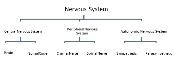
Central Nervous System
It comprises the brain and the spinal cord.
Brain
Brain accommodate in the skull while spinal cord is enclosed by the vertebral column. To support brain and protect it from external pressures, it is surrounded by a covering of triple membrane of connective tissue called the meninges. The three layers of meninges are duramater, arachnoid and piamater. The innermost layer, piamater is thin, delicate and highly vascular. It is firmly adhere to the brain. The middle layer is arachnoid membrane, which is thin and highly folded structure in front of cranial venous sinus. The villi like folding helps to reabsorb the cerebrospinal fluid (CSF). The outermost layer is very thick, strong and non elastic called duramatter, is made up of collagen fibre. The space between duramater and arachnoid is known as sub-dural space which is filled by serous fluid. The space between arachnoid and piamater is known as sub-arachnoid space and is filled by CSF.
CSF is lymph like clear and alkaline fluid whose function is to provide support as well as to protect the brain. It also helps in exchange of metabolic substances between the brain and blood capillaries. CSF mainly present in ventricle of brain, sub-arachnoid space and spinal cord.
The human brain is divided into three parts-
A. Fore brain or Prosencephalon: It includes cerebrum, diencephalon and olfactory lobes.
B. Mid brain or Mesencephalon: It consists of corpora quadrigemina and crura cerebri.
C. Hind brain or Rhombencephalon: It includes cerebellum, pons varolii and medulla oblongata.
A. Fore brain
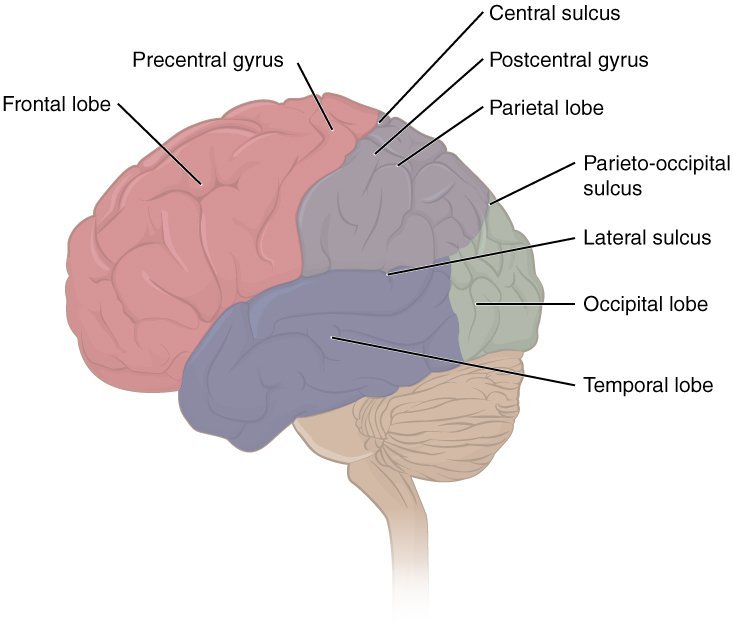
Cerebrum: It is the largest and most advanced part of brain which comprises of two cerebral hemispheres on the dorsal surface. A longitudinal groove is present between both cerebral hemisphere known as median fissure. Both hemispheres are somewhat connected with curved thick nerve fibres called corpus callosum. The outer portion of cerebrum is called the cerebral cortex which is made upcontains numerous cell bodies and relatively few myelinated axons. This gives as overall grey appearance and hence called asgrey matter. The surface of cortex is highly folded. The upward folds are called as gyri consecutive with the downward grooves called sulci. Beneath the grey matter there are millions of myelinated axon tracts and contains relatively very few cell bodies which give an opaque white appearance. Hence they are collectively called as white matter. Each cerebral hemisphere is divided into four lobes: frontal or anterior, parietal or middle, temporal or lateral and occipital or posterior lobe. Central sulcus separates frontal lobes from parietal lobes. Lateral sulcus or sylvian sulcus separate temporal lobe from frontal lobe and parietal lobe. Occipital lobe is separated by parietal lobe by parieto-occipital sulcus
There are three main functional area of each cerebral hemisphere.
1. Sensory area: They receive impulse form receptors.
2. Association area: They interpret the input, store the input and initiate a response. This area is involved in memory, learning and reasoning.
3. Motor area: They transmit impulse to the effector organs.
Diencephalon: It is small and posterior part of fore brain, covered by cerebrum. It consist of thalamus, hypothalamus, epithalamus and metathalamus.
i. Thalamus:It is a major part of diencephalon and represents upper lateral wall. It accepts all sensory impulses from the all part of body (except olfaction) and send those to the cerebral cortex. Thus it acts as relay centre. In lower animals thalamus act as sensory centre because cerebral cortex is less develop.
ii. Hypothalamus: It is called the master gland. It represents lower lateral wall of diencephalon. Pituitary gland is attached with its middle part. A web like structure is found on anterior surface of hypothalamus known as optic chiasma. In mammalian brain, corpus albicans is found on the posterior part of hypothalamus.
iii. Epithalamus: It represents the roof of diencephalon. Pineal gland is found in this region.
iv. Metathalamus: It represents the floor of diencephalon. It consist medial geniculate body (related to hearing) and lateral geniculate body (related to vision).
B. Mid Brain
It is small and contracted part of brain. Two longitudinal myelinated nerve fibres called cerebral penduclesor crura cerebrilocated at the anterior part of mid brain. Four spherical projections called optic lobe or colliculus are located at the posterior part of mid brain. Inferior optic lobes are related to acoustic reflex action.
C. Hind Brain
Cerebellum: It is second largest part of brain. Human cerebellum is made up of 3 lobes. Lateral lobes are large and spherical, called as cerebellar hemisphere. It control regulation and coordination of voluntary muscles.Cerebellumhelps to maintains the body balance of a person.
Medulla Oblongata: It is tubular and cylindrical in shape present at the posterior part of brain. It controls all the involuntary activities of the body e.g. respiration, metabolism, secretory actions of different cells etc.
Pons varolii: It is small spherical projection which is situated below the mid brain and upper to the medulla oblongata. It consists of many transverse and longitudinal nerve fibres.
Transverse nerve fibers are joined with cerebellum, whereas longitudinal fibre are join cerebrum to medulla oblongata. It regulates the breathing reaction through pneumotaxic centre.
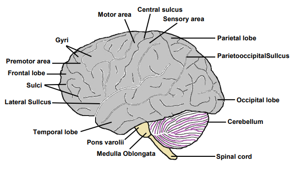
Midbrain, medulla oblongata and pons varolii are situated on one axis called brain stem. The side view of a human brain is shown in Figure which shows major parts of brain.
Name of Area | Location | Function |
Prefrontal cortex | Frontal lobe | Site of intelligence, knowledge and memory |
Premotor area | Frontal lobe | Writing centre, associated movement of eye, head & body, control complex movement of jaw, tongue, pharynx and larynx |
Motor area | Frontal lobe | Analysis of all type of voluntary muscle |
Frontal eye field | Frontal lobe | Opening and closing of eyelid and conjugate movement of eye |
Broca’s area or motor speech area | Frontal lobe | Analysis for speak |
Auditory area | Temporal lobe | Analysis for sound |
Olfactory area | Temporal lobe | Analysis for smell |
Wernicke’s area | Temporal lobe | Analysis for communication |
Gustatory area | Parietal | Analysis for taste |
Somesthetic area | Parietal | Analysis for touch, pain, pressure etc |
Angular gyrus | Parietal | Analysis for writing (associated with speech) |
Occipital area | Occipital | Analysis for vision |
2. Spinal Cord
Spinal cord is continuation of medulla oblongata which comes out from foramen of magnum and continues in neural canal of vertebral column. It is also covered by duramater, arachnoid and piamater. Narrow space between duramater and vertebra is known as epidural space. The outer part of spinal cord is of white matter while inner part is of grey matter. The grey matter projects outside and forms the dorsal and ventral horn. Dorsal and ventral horn continues in a tube like structure known as root of dorsal and ventral horn. In the root of dorsal horn, ganglia are present known as dorsal root ganglia. Sensory neurons are found in the dorsal root ganglia whose axon extend and get embedded into grey matter of spinal cord. These sensory neurons make synapse with ventral root neuron. Motor neurons are found in ventral root whose cyton is found in ventral horn while its dendrons are embedded into grey matter of spinal cord. Both sensory and motor nerve fiber combined and comes out from intervertebral foramen and form spinal nerve. Spinal cord acts as bridge between brain and organ of the body. It regulates and conducts the reflex action as well as it provides relay path for the impulses coming from brain.
Reflex Action
Reflex actions are involuntary actions, completed very quickly as compared to normal actions.
Reflex actions are of two types:
a. Cranial reflex: These actions are completed by brain.These are slow actions e.g. watering of mouth to see good food.
b. Spinal reflex: These actions are completed by spinal cord. These are fast actions e.g. withdrawal of arm at the time of pinching by any needle
Peripheral Nervous System
All nerves arise from brain and spinal cord are included in peripheral nervous system. Nerves arises from brain is termed as cranial nerve whereas nerves arises from spinal cord is termed as spinal nerves. All reptiles, birds and mammals have 12 pairs of cranial nerve. Amphibians and fishes have only 10 pairs of cranial nerves. In human, I, II, and VIII cranial nerve are pure sensory in nature. III, IV, VI, XI and XII cranial nerve are motor nerve and rest others out of 12 cranial nerves are mixed type of nerves.
Name | Origin | Distribution | Nature | Function |
Olfactory | Olfactory epithelium | From olfactory lobe to temporal lobe | Sensory | Smell |
Optic | Retina | Leads to occipital lobe | Sensory | Sight |
Oculomotor | Midbrain | Four eye muscle | Motor | Movement of eyeball |
Trochlear | Midbrain | Superior oblique eye muscle | Motor | Rotation of eyeball |
Trigeminal | Pons varolii | Skin of nose, eyelid, forehead, scalpe, conjunctiva, lachrymal gland. Mucous membrane of cheeks and upper lip and lower eyelid Lower jaw, lower lip, pinna | Mixed
|
|
a. Ophthalmic | Sensory | Sensory supply to concerning part | ||
b. Maxillary | Sensory | – | ||
c. Mandibular | Mixed
| Muscle of mastication | ||
Abducens | Pons varolii | Lateral rectus eye muscle | Motor | Rotation of eyeball |
Facial | Pons varolii | Face, neck, taste buds, salivary gland | Mixed | Taste (anterior 2/3 part of tonge), facial expression, saliva secretion |
Auditory | Pons varolii | Internal ear | Sensory | Hearing and equilibrium |
Glossopharyngeal | Medulla oblongata | Muscle and mucous membrane of pharynx and tongue. | Mixed | Taste (posterior 1/3 part of tounge), saliva secretion |
Vagus | Medulla oblongata | Larynx, lungs. Heart, stomach, intestines | Mixed | Visceral sensations and movements |
Accessory spinal | Medulla oblongata | Muscles of pharynx and larynx | Motor | Movement of pharynx and larynx |
Hypoglossal | Medulla oblongata | Muscles of tongue | Motor | Movement of tounge. |
In human, there are 31 pairs of spinal nerves. Each spinal nerve is of mixed type and arise
from the roots of the horns of grey matter of the spinal cord.
Spinal nerves are divided into 5 groups according to its position:
- Cranial spinal nerve – 8 pairs
- Thoracic spinal nerve – 12 pair
- Lumber spinal nerve – 5 pairs
- Sacral spinal nerve – 5 pairs
- Coccygeal nerve – 1 pairs
Autonomic nervous system
The autonomic nervous system controls activities inside the body that are involuntary e.g. heart rate, sweating, peristalsis etc. It consists of motor neurons passing to the smooth muscle of internal organs. Autonomic nervous system plays an important role in maintaining homeostasis. It is divided into two parts: 1) Sympathetic and 2) Parasympathetic.
Sympathetic system is related with such intuitive reaction which increases the protection of body in adverse atmospheric condition along with energy consumption. Whereas parasympathetic system is linked with those reactions in which energy is conserved.
Measurement of nerve conduction
A nerve conduction study (NCS) is a medical diagnostic test. It is used to estimate the function and the capability of electrical conduction, of the motor nerves and sensory nerves of the human body. Nerve conduction velocity (NCV) is frequently measured during this test.
NCS laterally with electromyography measure nerve and muscle function. Diagnosis of defective spinal nerve compression, or any other neurologic disorder or injuries are undertaken for study by NCS process. Evaluation of numbness of limbs, weakness of the legs and arms as well tingling or burning sensation in certain areas of the body are the central area of diagnosis.. Nerve conduction study mainly comprise of the following studies.
- Motor NCS: It is performed by electrical stimulation of a peripheral nerve and recording from a muscle to which these nerve supplies. Latency is defined as the time taken for the electrical impulse to travel from the stimulation to that muscleand is usually measured in milliseconds. The target muscle generates a response whose size is called the amplitude. Motor amplitudes are measured in millivolts. Determination of NCV across different segments of the nerve is a primary goal of the Motor NCS study. It is done by the stimulation of two or more different locations along the same nerve.With the help of the difference in latencies from the two points of stimulation as well as the distance between the different stimulating electrodes, one can calculate the NCVs across different segments
- Sensory NCS: It is similar to Motor NCS and is performed by electrical stimulation of a peripheral nerve and but here the recording is done from a truly sensory portion of the nerve. Sensory latencies are measured milliseconds but sensory amplitudes are much smaller than the motor amplitudes (microvolt). Sensory NCV is calculated in the same way as motor NCVs
- F-wave study: The motor and sensory segments are concerned about nerve conduction velocities in sections/segments of limb whereas the F-wave latency is used to derive the conduction velocity of nerve between the limb and spine.In a typical F wave study, a strong electrical stimulus is applied above the distal portion of a nerve. The resulting the impulse travels both in both directions: one towards the muscle fibre and the other back to the motor neurons of the spinal cord. These directions are referred to as orthodromic and antidromic, respectively. The orthodromic stimulus on reaching the muscle fibre elicits a strong Mresponse indicative of muscle contraction. Meanwhile the antidromic stimulus on reaching the motor neuron cell bodies excites a small portion of the motor neurons causing them to backfire resulting in orthodromic wave which travels back down the nerve towards the muscle. This reflected stimulus evokes a smaller response of the muscle fibres resultingin a second CMAP called the F wave. The limb length, D(in millimetres)is taken into account for calculations of Conduction velocity.
- H-reflex study: This studyis similar to F-wave study and evaluates conduction between the limb and the spinal cord. Although a subtle difference exists in that here the impulses going toward the spinal cord are in sensory nerves while the impulses coming from the spinal cord are in motor nerves
The interpretation of nerve conduction studies is a complex affair and expert medical practitioners such as neurologists, physiatrists or clinical neurophysiologists are routinely involved. NCS have proven to be very helpful in diagnosis of many diseases related to the nerves. The process is non-invasive, albeit sometimes it can be painful due to minor electrical shocks. Although the low amount of electrical current is considered safe, patients with a harbouring electrical devices such as permanent pacemaker are advised to avoid this kind of test or tell the examiner prior to the test.
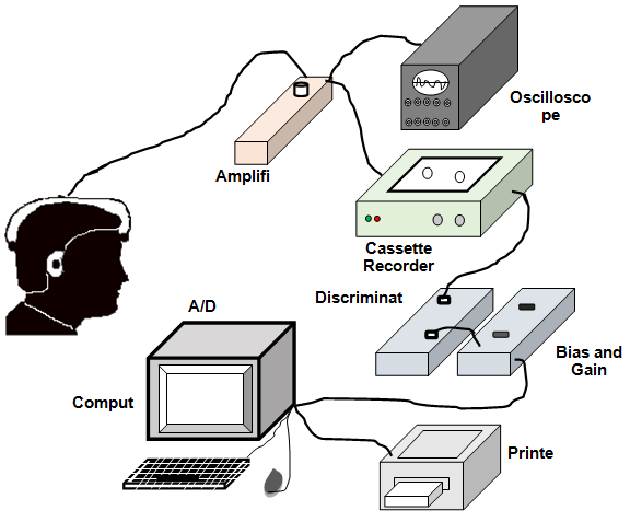
Electro-encephalograph (EEG)
In 1929, Hens Berger is credited to have found some electrical activity when he connect a galvanometer to human scalp. It gave birth to electro-encephalography. EEG is an electrically operated instrument having array of 16-30 electrode, which when attached to the scalp for short time gives electric wave signals. It operates by detecting the brain’s electrical charge which is maintained by billions of neurons. The Neurons are electrically charged due to continuous pumping of ions by membrane transport proteins across their membrane. Neurons constantly exchange ions with the extracellular fluid, e.g. to maintain resting membrane potential. When many ions having similar charge are pushed out of several neurons at the same time, they can push their neighbours, who further apply force to their neighbours, and so on such that a wave forms. When the wave of ions reaches the electrodes attached to the scalp, they can give or take electrons on or from the metal of the electrodes. Since metal can conducts these electrons easily, voltages difference between any two electrodes can be measured by a voltmeter. Recording these voltages for a specific time gives us the EEG. A schematic of typical EEG frequency display system is shown in Figure.
Wave patterns form during EEG recording
Delta waves: The frequency of Delta wave is below 4 Hz. It is the highest in amplitude and the slowest waves. It originate normally in adults during sleep. It is also seen normally in babies. It can also be observed in patients during coma.
Theta waves: The frequency of theta wave range from 4 Hz to 7 Hz. It is seen normally in young children. It may be seen in older children and adults under stress or during meditation. Excess theta for age represents abnormal activity.
Alpha waves: The frequency of alpha wave range from 7 Hz to 14 Hz. An awake but resting person produces alpha wave. Hans Berger termed “alpha wave” when he saw the first rhythmic EEG activity. This was the “posterior basic rhythm” seen in the occipital regions of the brain. It arises with the closing of the eyes and with relaxation and weakens with eye opening or mental labour. In addition to the posterior basic rhythm, there are other normal alpha rhythms such as the “mu rhythm” which arises when the hands and arms are indolent.
Beta waves: The frequency of beta wave range from 15 Hz to about 30 Hz. During extreme mental activity, beta wave initiates from frontal and parietal regions. Beta activity is closely linked to motor behaviour and is generally weakened during active movements. An alert wide awake person shows unsynchronised beta wave.
Gamma waves: The frequency of gamma wave is nearly 30–100 Hz. Gamma rhythms represent binding of different populations of neurons together into a network for the purpose of carrying a certain motor function.
Mu waves: The frequency of mu wave is 8–13 Hz. It partly overlaps with other frequencies. It denotes the synchronous firing of motor neurons in rest state. Deviations from normal pattern indicate brain disorder and change in physiological state of brain. EEG can diagnose epilepsy, brain tumour, abscess, sleep disorders, metabolic and drug effects on brain.


It is very good to learn .