Landmarks in the study of a cell
Soon after Anton Van Leeuwenhoek invented the microscope, Robert Hooke in 1665 observed a piece of cork under the microscope and found it to be made of small compartments which he called “cells” (Latin cell = small room). In 1672, Leeuwenhoek observed bacteria, sperms and red blood corpuscles, all of which were cells. Much later, in 1831, Robert Brown, an Englishman observed that all cells had a centrally positioned body which he termed the nucleus.
The cell theory
In 1838 M.J. Schleiden and Theodore Schwann formulated the “cell theory.” Which maintains that:
- all organisms are composed of cells.
- cell is the structural and functional unit of life, and
- cells arise from pre-existing cells.
The cells vary considerably, in shapes and sizes. Nerve cells of animals have long extensions. They can be several centimetres in length. Muscle cells are elongated in shape. Egg of the ostrich is the largest cell (75 mm). Some plant cells have thick walls. There is also wide variation in the number of cells in different organisms.
The Cell
A cell may be defined as a unit of protoplasm bound by a plasma or cell membrane and possessing a nucleus. Protoplasm is the life giving substance and includes the cytoplasm and the nucleus. The cytoplasm has in it organelles such as ribosomes, mitochondria, golgi bodies, plastids, lysosomes and endoplasmic reticulum. Plant cells have in their cytoplasm, large vacuoles containing non-living inclusions like crystals, and pigments. The bacteria have neither defined cell organelles nor a well formed nucleus. But every cell has three major components:
- plasma membrane
- cytoplasm
- DNA (naked in bacteria) and enclosed by a nuclear membrane in all other organisms
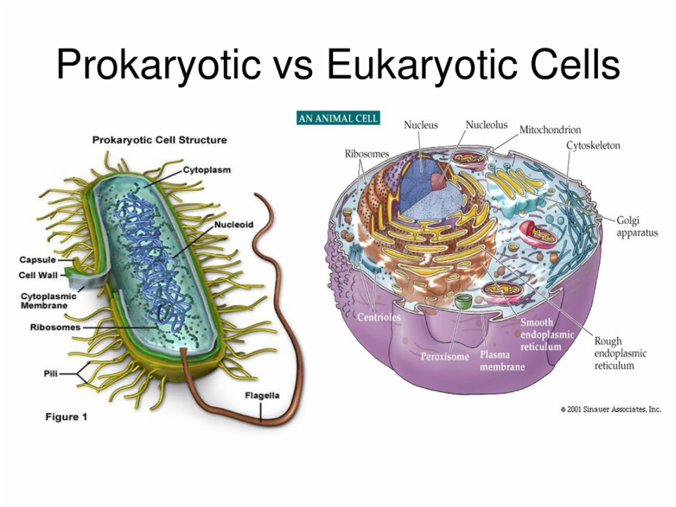
Two basic types of cells
Cytologists recognize two basic types of cells (Fig. 4.1). Their differences have been tabulated below in Table. Organisms which do not possess a well-formed nucleus are prokaryotes such as the bacteria. All others possess a well-defined nucleus, covered by a nuclear membrane. They are eukaryotes.
Eukaryotic cell (eu = true, karyon = nucleus) | Prokaryotic cell (Pro = early/primitive) |
1. Nucleus distinct, with a well-formed nuclear membrane. | 1. Nucleus not distinct, it is in the form of a nuclear zone ‘nucleoid’. Nuclear membrane absent. |
2. Double-membraned cell organelles (Chloroplasts, mitochondria, nucleus) and single membraned (Golgi apparatus, lysosomes, vacuole, endoplasm reticulum) are present | 2. Single-membraned cell bodies like mesosomes present. Endoplasmic reticulum, plastids, nitochondria microbodies like lysosomes, and Golgi body absent. |
3. Ribosomes – 80 S | 3. Ribosomes – 70 S |
4. Distinct compartments in the cell i.e. the cytoplasm and the nucleus | 4. No compartments. |
5. Depending upon the species number of chromosomes per nucleus varies from two to many. | 5. There is only one chromosome per cell |
6. Each chromosome is linear with its two ends free. | 6. The chromosome is circular and remains attached to cell membrane at one point. |
7. Each chromosome has one linear double-stranded DNA complexed with histones | 7. The chromosome has single double-stranded circular DNA molecule and is not associated with histones. |
8. Each chromosome has one centromere that divides a chromosome into two arms. However, if the centromere is terminal, the chromosome would have only one arm | 8. The chromosome lacks a centromere |

Svedberg unit
When the cell is fractionated or broken down into its components by rotating
in an ultracentrifuge at different speeds the ribosomes of eukaryotic and
prokaryotic cells sediment (settle down) at different speeds. The coefficient of
sedimentation is represented in Svedberg unit and is depicted as S.
The plant cell and the animal cell also differ in several respects as given in Table
Plant cell | Animal cell |
| 1. Cellulose cell wall present external to cell membrane. | 1. No cell wall, outermost structure is cell membrane or plasma membrane |
| 2. Vacuoles are usually large. | 2. Generally vacuoles are absent and if present, are usually small. |
| 3. Plastids present | 3. Plastids absent. |
| 4. Golgi body present in the form of units known as dictyosomes. | 4. Golgi body well developed having 2 cisternae |
| 5. Centriole absent. | 5. Centriole present. |


Leave a Reply
You must be logged in to post a comment.