Exploring the Human Eye: Unveiling its Anatomy, Parts, and Structure
The human eye is a marvel of complexity and precision, allowing us to perceive the vibrant world around us. From the simplest forms of light detection to the intricate details of color and depth perception, the eye plays a pivotal role in our sensory experience. In this article, we’ll delve into the fascinating anatomy, parts, and structure of the human eye.
Anatomy of the Eye
The eye is a globe-shaped organ nestled within the eye socket (orbit) of the skull. It consists of several layers that work together to capture and process light.
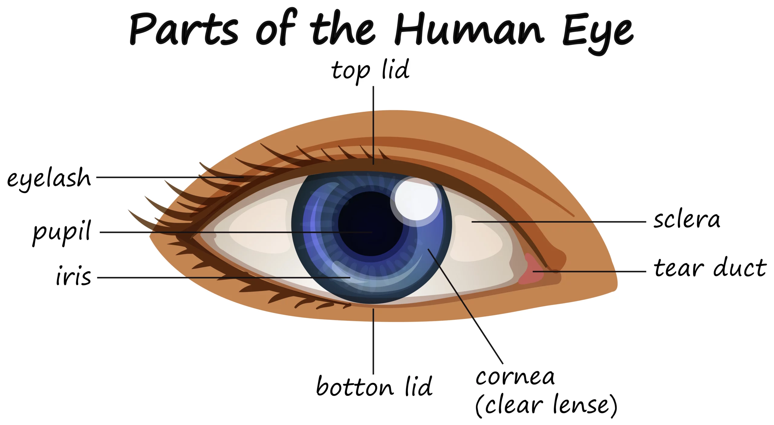
Sclera: The outermost layer is the sclera, commonly known as the “white of the eye.” It’s a tough and protective layer that maintains the eye’s shape.
Cornea: The transparent cornea covers the front portion of the eye. It’s responsible for bending light rays and focusing them onto the retina.
Iris: The colored part of the eye is the iris. It controls the size of the pupil, the central black hole that allows light to enter the eye.
Pupil: As mentioned, the pupil is the central aperture of the eye. It adjusts its size to regulate the amount of light entering the eye.
Lens: Behind the iris, the lens further refracts light and fine-tunes its focus on the retina. The lens can change shape to focus on objects at different distances, a process known as accommodation.
Retina: The innermost layer is the retina, a thin, light-sensitive tissue akin to the film in a camera. It contains specialized cells called photoreceptors (rods and cones) that convert light into electrical signals.
Parts of the Eye
Now, let’s take a closer look at the key components within the eye:
Optic Nerve: The optic nerve connects the retina to the brain, transmitting the electrical signals generated by photoreceptors to the brain for processing.
Aqueous Humor and Vitreous Humor: The eye contains two types of fluid: the aqueous humor, which fills the space between the cornea and lens, and the vitreous humor, a jelly-like substance that fills the space behind the lens. These fluids maintain the eye’s shape and provide nutrients to its cells.
Ciliary Muscles: These muscles surround the lens and help it change shape for focusing on objects at various distances.
Choroid: The choroid is a layer between the retina and the sclera that supplies blood to the retina and helps prevent light from scattering within the eye.
Conjunctiva: This thin, transparent membrane covers the front of the eye and lines the inside of the eyelids, helping to protect the eye and keep it moist.
Structure of Vision
The process of vision can be broken down into these steps:
Light Entry: Light enters the eye through the cornea and pupil.
Focusing: The cornea and lens focus the light onto the retina.
Photoreception: Photoreceptor cells in the retina (rods and cones) detect the light and convert it into electrical signals.
Signal Transmission: The electrical signals travel through the optic nerve to the brain.
Brain Interpretation: The brain processes the signals to create the images we perceive, including colors, shapes, and depth.
The human eye is an extraordinary sensory organ with a complex structure that allows us to experience the world in vivid detail. From its protective outer layers to the intricate mechanisms for focusing and processing light, the eye’s anatomy and parts work in harmony to grant us the gift of vision.