Which organelle divides and compartmentalizes the cell?
The endomembrane system, which fills the cell and divides it into compartments, and is visible only through electron microscopy. The endoplasmic reticulum (ER), a series of interconnected membranous tubes and channels in the cytoplasm, is the largest and most extensive system of the internal membranes.
What is the function of the endomembrane system?
The endomembrane system allows macromolecules to diffuse or be transferred from one of the components of the system to another.
What is the difference between rough and smooth endoplasmic reticulum?
Rough endoplasmic reticulum are regions rich in ribosomes that manufacture proteins. These regions appear to have a pebbly surface that is somewhat similar to sand-paper. Regions with no ribosomes are referred to as smooth endoplasmic reticulum. The smooth endoplasmic reticulum is involved in the synthesis of lipids and steroids, carbohydrate metabolism, and the inactivation and detoxification of drugs and other components that may be toxic or harmful to the cell.
What is the function of the Golgi apparatus?
The Golgi apparatus (frequently called the Golgi body) is a collection of flattened stacks of membranes. It serves as the packaging centre for cell products. It collects materials at one place in the cell, and packages them into vesicles for use elsewhere in the cell or transportation out of the cell.
Who discovered the Golgi apparatus?
In 1898 Camillo Golgi (1843–1926), an Italian physician, first described an irregular network of small fibers, cavities, and granules in nerve cells. It was not until the 1940s, and the invention of the electron microscope, that the existence of the Golgi apparatus was confirmed. In 1906 Golgi and Santiago Ramón y Cajal (1852–1934) were awarded the Nobel Prize for Physiology or Medicine for their investigations into the fine structure of the nervous system.
How many Golgi bodies are in a cell?
Protists contain one or a small number of Golgi bodies. Animal cells may contain twenty or more Golgi bodies, while plant cells may contain several hundred Golgi bodies.
What are the two major types of vesicles in eukaryotic cells?
Vesicles are small, intracellular membrane sacs usually formed from the Golgi apparatus. Lysosomes and microbodies are examples of vesicles found in eukaryotic cells.
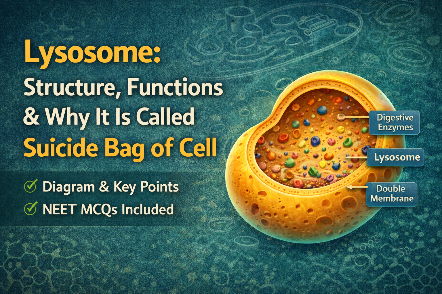
What is the function of lysosomes?
Lysosomes are single, membrane-bound sacs that contain digestive enzymes. The digestive enzymes break down all the major classes of macromolecules including proteins, carbohydrates, fats, and nucleic acids. Throughout a cell’s lifetime, the lysosomal enzymes digest old organelles to make room for newly formed organelles. The lysosomes allow cells to continually renew themselves and prevent the accumulation of cellular toxins.
Who discovered lysosomes?
Lysosomes are a relatively modern discovery in cell biology; they were observed by 42 Christian de Duve (1917– )in the early 1950s. In 1955, after six years of experiments, de Duve was convinced that he had found an organelle that had not been previously described and was involved in intracellular lysis (digestion). He named the organelle a lysosome. This organelle was the first to be described entirely on biochemical criteria. The results were later verified using electron microscopy. In 1974 De Duve, Albert Claude (1898–1983), and George Palade (1912–) shared the Nobel Prize in Physiology or Medicine for their work detailing the functions of the lysosome.
What is a peroxisome and what is its function in a cell?
Peroxisomes, also discovered by Christian de Duve (1917–), are surrounded by a single membrane and are the most common type of microbody in cells. They are especially prominent in algae, the photosynthetic cells of plants, and both mammalian kidney and liver cells. Peroxisomes contain detoxifying enzymes and produce catalase, which breaks down hydrogen peroxide into hydrogen and water.
Why are ribosomes an important organelle?
Ribosomes, one of the most complex aspects of the molecular machine, are the site of protein synthesis in a cell. They consist of a large and small subunit composed of ribosomal RNA and protein. However, compared with membrane-bound organelles, ribosomes are tiny structures!
How many ribosomes are in a typical cell?
A bacterial cell will typically have a few thousand ribosomes, while a human liver cell contains several million ribosomes. Actively growing mammalian cells contain five to ten million ribosomes that must be synthesized each time the cell divides.
Where are ribosomes found in a cell?
Ribosomes are found in the cytoplasm of both prokaryotic and eukaryotic cells, as well as in the matrix of mitochondria and the stroma of chloroplasts. In eukaryotic cytoplasm, ribosomes are found in the cystol, and are bound to the endoplasmic reticulum as well as the outer membrane of the nuclear envelope.
How are ribosomes different from other organelles?
Ribosomes differ from most other organelles in not being bound by a membrane.
Are there differences between prokaryotic and eukaryotic ribosomes?
Prokaryotic and eukaryotic ribosomes resemble each other structurally, but they are not identical. Prokaryotic ribosomes are smaller in size, contain fewer proteins, have smaller RNA molecules, and are sensitive to different inhibitors of protein synthesis. What are mitochondria? A mitochondrion (singular) is a self-replicating, double-membraned body found in the cytoplasm of all eukaryotic cells. The outer membrane of a mitochondrion is smooth, while the inner membrane is folded into numerous layers that are called cristae. Mitochondria are the location for much of the metabolism necessary for protein synthesis, and the production of both ATP and lipids.
When were mitochondria discovered?
In 1857 Rudolf Albert von Kölliker (1817–1905), histologist and embryologist, first described “sarcosomes” (now called mitochondria) in muscle cells. The term “mitochondrion” (meaning “threadlike granule”) was first used in 1898. Functionally active mitochondria were first isolated in 1948. Kölliker was among the first biologists to interpret tissue structure in terms of cellular elements.
How many mitochondria are there in a cell?
The number of mitochondria varies according to the type of cell. The number ranges 44 between one and 10,000, but averages about 200. Each cell in the human liver has over 1,000 mitochondria. Cells with high energy requirements, such as muscle cells, may have many more mitochondria.
What is the function of chloroplasts?
Chloroplasts are the structural and functional units where photosynthesis takes place—the process whereby green plants use light energy for the synthesis of organic molecules from carbon dioxide and water, with oxygen released as a by-product. They contain the green pigment chlorophyll, which traps light energy for photosynthesis. When were chloroplasts first described? Because chloroplasts are a large cell structure (bigger than any other organelle except the nucleus), they were described and studied early in the history of cytology. Anton van Leeuwenhoek (1632–1723) and Nehemiah Grew (1641–1712) described these organelles in the seventeenth century, in the early stages of the microscopic study of cells.
Do all plant cells contain chloroplasts?
No, not all plant cells contain chloroplasts. The various types of plant cells arise from meristem, rapidly dividing and undifferentiated tissue. Meristem cells do not contain chloroplasts, but have smaller organelles called proplastids. Depending on their location in a plant and how much light they receive, proplastids develop into one of several kinds of plastids with different functions. Chloroplasts are one example of a plastid that converts light energy to chemical energy, subsequently used in synthesizing organic molecules—the process of photosynthesis.
What are the different types of plastids?
Proplastids can differentiate into several types of plastids that are involved in cellular storage. Amyloplasts store starch, proteinoplasts store proteins, and elaioplasts store lipids. In addition, some proplastids develop into chromoplasts.
What are the main components of chloroplasts?
Chloroplasts have outer and inner membranes, which are in close association with each other. They also have a closed compartment of stacked membranes—called 46 grana—that lie inside the inner membrane. A chloroplast may contain one hundred or more grana, and each granum may contain a few to several dozen disk-shaped structures called thylakoids that contain chlorophyll on their surface. The fluid that surrounds the thylakoid is called the stroma.
How many chloroplasts are in plant cells?
Unicellular algae may only have one large chloroplast, whereas a plant leaf cell may have between 20 and 100.
How do chromoplasts differ from chloroplasts?
Chromoplasts are pigment-containing plastids found in plants. They are responsible for the characteristic red, orange, or yellow coloration visible in some flowers, fruits, and plants.
What is the cytoskeleton and what is its function?
The cytoskeleton is a structural feature of eukaryotic cells that was revealed by advanced microscopy. It consists of an extensive three-dimensional network of interconnected filaments and tubules that extends throughout the cytosol, from the nucleus to the inner surface of the cell membrane. These filaments and tubules determine cell shape and facilitate a variety of cell movements.
What are the three types of fibers found in the cytoskeleton of eukaryotic cells?
The cytoskeleton is a network of fibers that maintains the shape of cells. The three types of fibers are actin filaments, microtubules, and intermediate filaments. Actin filaments are long fibers composed of two protein chains. They are responsible for cellular movements, such as contraction, crawling, “pinching” during division, and formation of cellular extensions. Microtubules are hollow tubes composed of a ring of thirteen protein filaments. They are responsible for moving materials within the cell. Intermediate filaments are tough, fibrous protein molecules structured in an overlapping arrangement. They are intermediate in size when compared to actin filaments and microtubules, and provide structural stability to cells.