Understanding Oxygenation Challenges in the Heart
Ventricular Septal Defect (VSD) is one of the most common congenital heart defects found in infants. It affects how oxygen-rich and oxygen-poor blood mix inside the heart, leading to potential complications in oxygen delivery to the body.
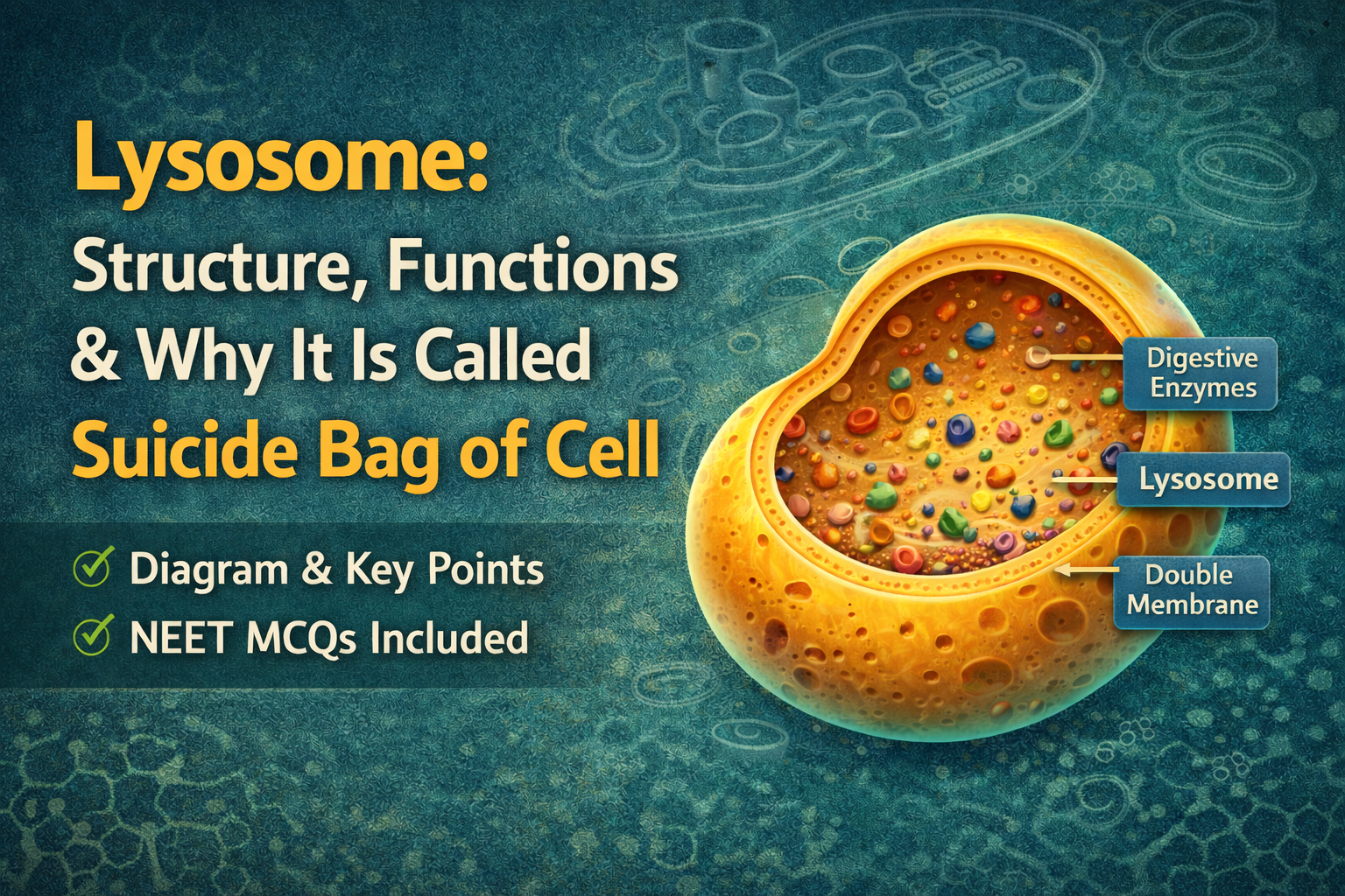
Image by Freepik
Quick Overview
- VSD is a hole in the wall (septum) between two ventricles
- Causes mixing of oxygenated and deoxygenated blood
- Can lead to breathing difficulty in infants
- Small VSD may close naturally
- Severe cases require medical or surgical treatment
What is Ventricular Septal Defect?
Ventricular Septal Defect (VSD) is a condition where there is an abnormal opening in the septum separating the left and right ventricles of the heart.
This opening allows oxygen-rich blood from the left ventricle to mix with oxygen-poor blood in the right ventricle.
How VSD Affects Oxygenation in Infants
In a normal heart:
- Oxygen-rich blood flows from lungs → left side → body
In VSD:
- Blood flows from left ventricle → right ventricle
- This causes excess blood to go to lungs
- Less efficient oxygen delivery to body
Result:
- Increased workload on heart
- Poor oxygen supply in severe cases
Types of Ventricular Septal Defect
- Perimembranous VSD (most common)
- Muscular VSD
- Inlet VSD
- Outlet VSD
Symptoms of VSD in Infants
- Rapid breathing
- Poor feeding
- Sweating while feeding
- Slow weight gain
- Fatigue
Causes of VSD
- Congenital (present at birth)
- Genetic factors
- Abnormal heart development during pregnancy
How is VSD Diagnosed?
- Physical examination (heart murmur)
- Echocardiogram (main test)
- Chest X-ray
- ECG
Treatment Options for VSD
- Small VSD: may close naturally
- Medications: to manage symptoms
- Surgery: for large defects
- Catheter-based repair
Possible Complications
- Heart failure
- Pulmonary hypertension
- Delayed growth
Conclusion
Ventricular Septal Defect is a common but manageable condition in infants. Early diagnosis and proper treatment help ensure normal growth and development.